FU Jie, GAO Zhibing, JIA Hai, LIU Siqi, LI Huabai, ZHANG Long, LI Zhilin, ZHAO Chenguang
2026, 35(2): 149-161.
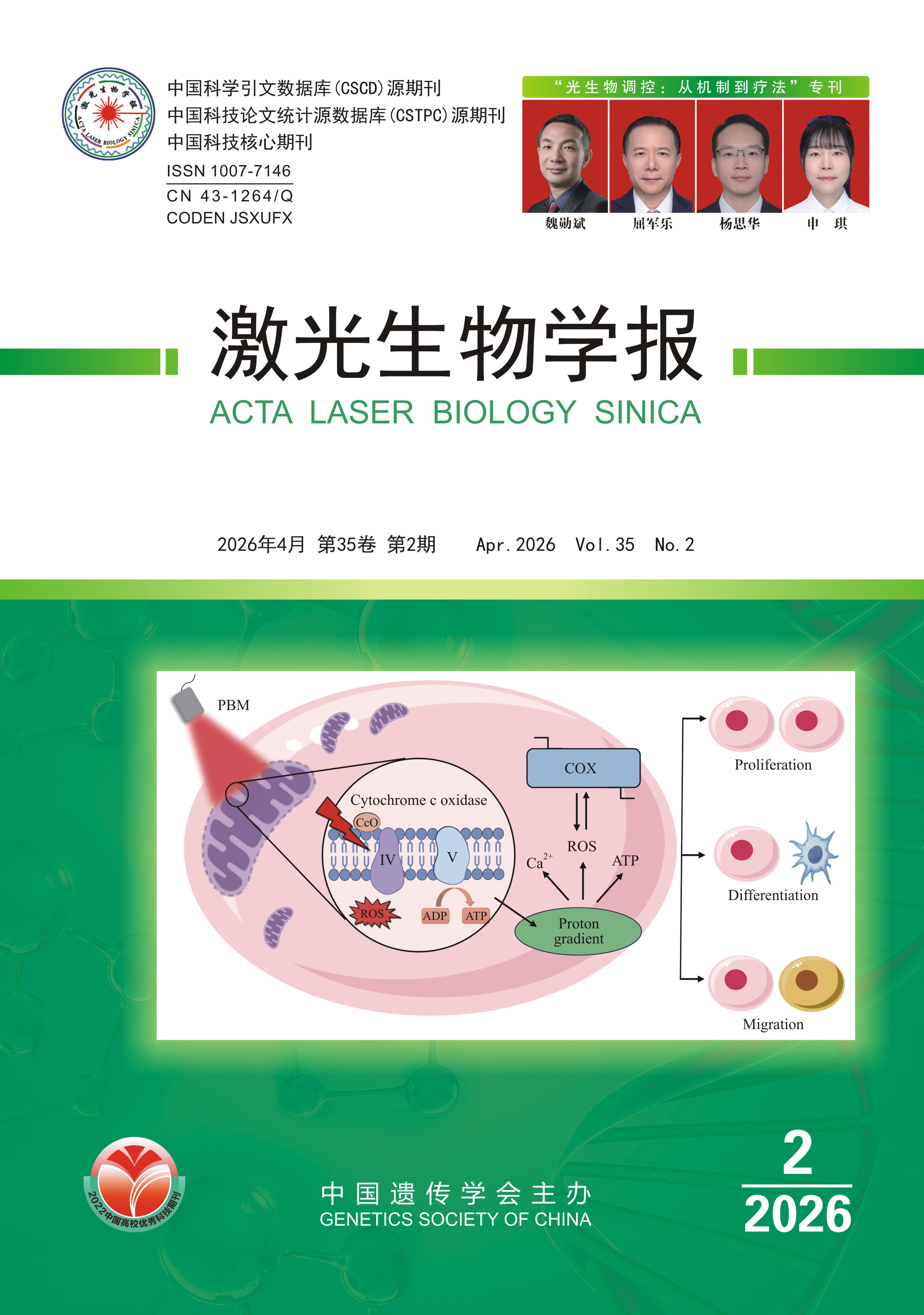
Abstract: As a complex neurodegenerative disorder, Alzheimer’s disease (AD) urgently requires multi-targeted and personalized treatment strategies. Transcranial photobiomodulation (tPBM), a non-invasive neuromodulation technique, utilizes near-infrared light to penetrate the cranium and to target multiple pathological processes, such as mitochondrial dysfunction, neurovascular unit impairment, and glymphatic system dysregulation. Possessing dual properties of metabolic regulation and neuromodulation, tPBM demonstrates significant potential for multi-target intervention, aligning with mainstream drug development strategies, e.g., amyloid β-protein (Aβ) clearance and inflammation modulation. This review provides an analysis of 42 included clinical studies, specifically focusing on sample characteristics, intervention parameters and clinical outcomes, and comprehensively synthesizes mechanistic evidence, clinical advancements, and dosing dosimetric methods of tPBM in AD intervention, emphasizing its applicability in home-based and wearable device trends, while proposing a translational framework that links transcranial optical dosimetry with biological effects. Future advancements may enable tPBM to evolve from population-wide effectiveness to individualized therapy through constructing personalized photon dose-response models integrated with real-time physiological feedback and brain-imaging navigation, thereby positioning tPBM as a pivotal non-pharmacological intervention for AD synergistic treatment and long-term management.
Key words: Alzheimer’s disease; transcranial photobiomodulation; dosimetry; multitarget therapy; neuromodulation
(Acta Laser Biology Sinica, 2026, 35(2): 149-161)
